
Case Studies
MiraTyper on Real Tissue
Examples and evaluations across H&E, single-cell, and spatial datasets, with the measurements and reference data shown alongside the results.
Colon Cancer • H&E
Linking H&E Tissue Patterns to Microsatellite Instability in Colon Cancer
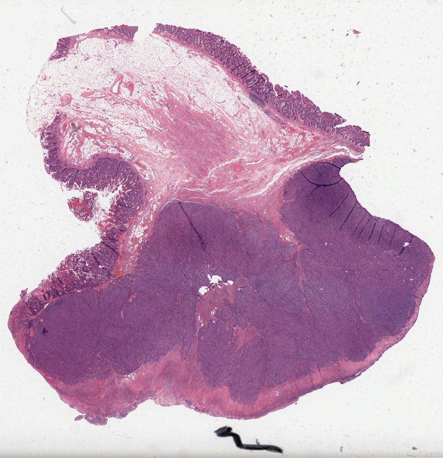
121–128
Slides, by Analysis
0.760
Spatial-model AUC
24 µm
Tile Width
Cervical Cancer • Spatial Transcriptomics
Density, not Triad Geometry, drives Allograft Rejection Signature in Reference Cervical Cancer Slide
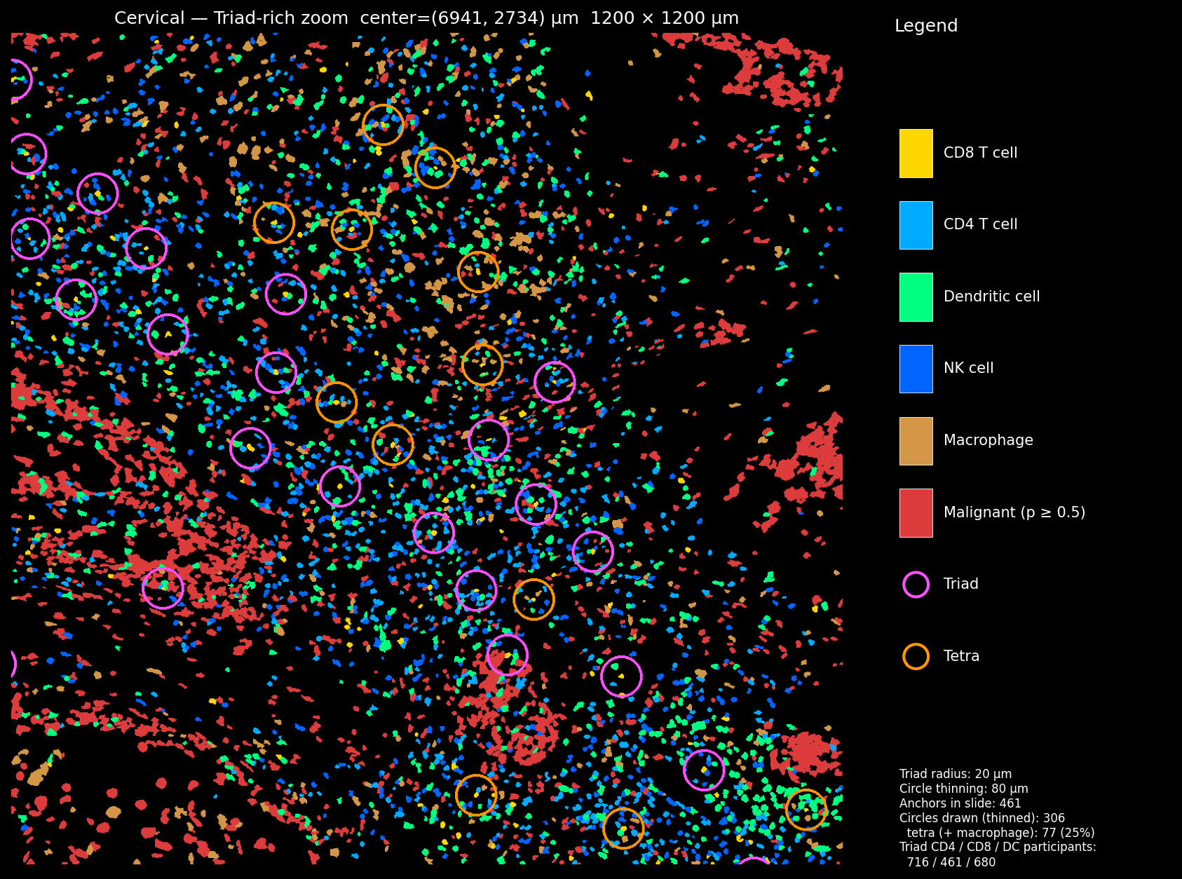
717,576
Cells Analyzed
406
Triad-Anchor CD8 Cells
11.6%
Mature-TLS Fraction
Lymph Node • Spatial Transcriptomics
MiraTyper Evaluation on Xenium 5K Human Lymph Node (708K Cells)
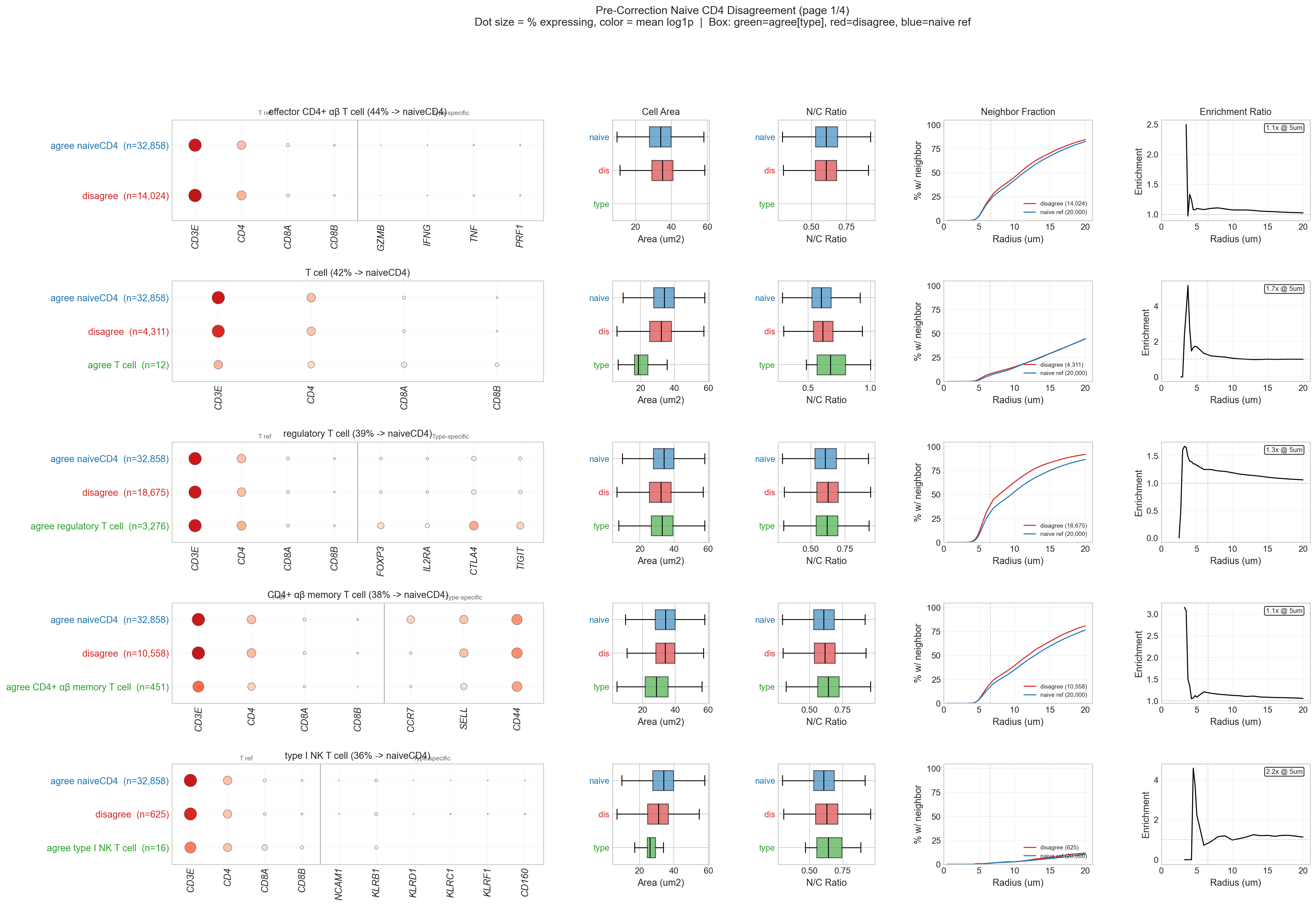
708,983
Cells Evaluated
0.813
Corrected h-F1
76.5%
Better Spatial Coherence
Pancreas • Type 2 Diabetes
MiraTyper Performance on Pancreatic Type 2 Diabetes Single-Cell Datasets
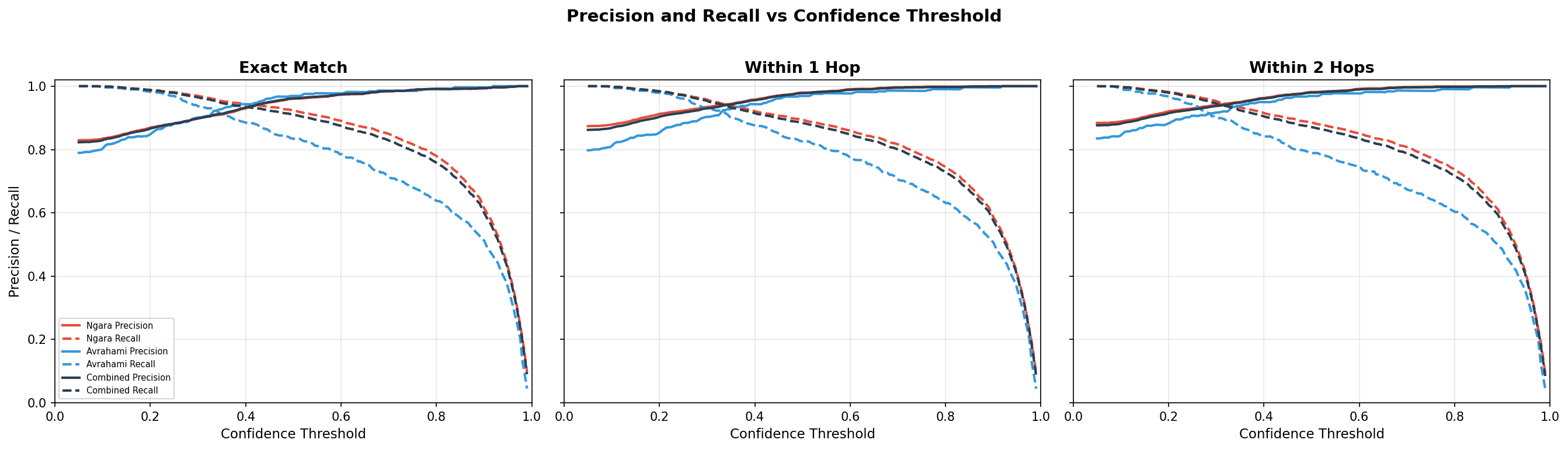
3,974
Cells Evaluated
96.2%
Accuracy (conf ≥ 0.5)
600
Annotation Corrections
More Case Studies Coming Soon
We're continually evaluating MiraTyper on new tissues and platforms.
Want to See MiraTyper on Your Data?
Request a demo and we'll show you MiraTyper running on a dataset like yours.